Recreate renal physiology with microfluidic kidney-on-a-chip systems. Exposing primary proximal tubule cells to fluidic shear stress restores key transporter expressions and cellular polarity, facilitating the assessment of renal drug excretion, reabsorption, and drug-induced kidney injury (DIKI).
- Introduction
- Service Details
- Workflow
- Features
- FAQs
Microphysiological Renal Models for Clearance Studies
The kidney is a primary route for drug elimination and a major target for drug-induced toxicity. However, standard static cultures of renal proximal tubule epithelial cells (RPTECs) lack tubular fluid flow, which typically leads to a loss of columnar polarity and the rapid downregulation of key drug transporters (e.g., OATs and OCTs).
Creative Bioarray’s Kidney-on-a-Chip platform incorporates controlled microfluidic shear stress to simulate the human nephron microenvironment. This mechanical cue restores the expression of functional influx and efflux transporters and recreates the renal barrier, enabling the quantitative assessment of active tubular secretion and nephrotoxic liabilities.
Kidney Clearance & Toxicity Services
1. Renal Clearance & Active Secretion Services
- Differentiation between glomerular filtration, active tubular secretion, and reabsorption.
- Assessment of drug excretion kinetics across the polarized epithelial barrier.
- Prediction of total renal clearance (CLR) for small molecules and biologics.
2. Transporter-Mediated DDI Services
- Quantification of substrate transport involving basolateral uptake (OAT1, OAT3, OCT2).
- Evaluation of apical efflux mechanisms via MATE1, MATE2-K, P-gp, and MRPs.
- Identification of specific inhibitors to determine renal Drug-Drug Interaction (DDI) risks.
3. Drug-Induced Kidney Injury (DIKI) Services
- Detection of early nephrotoxicity biomarkers (KIM-1, NGAL, Clusterin) released directly into the apical effluent.
- Assessment of mitochondrial toxicity and intracellular drug accumulation (e.g., Aminoglycosides, Cisplatin).
Analytical Endpoints and Platform Specifications
Instrumentation:
- Programmable shear stress controllers
- Confocal microscopy for 3D z-stacking
Key Reagents:
- Validated primary human RPTECs
- Fluorescent transporter substrates
Analysis Methods:
- ELISA for toxicity biomarkers (KIM-1/NGAL)
- Intracellular accumulation assays
- Trans-epithelial electrical resistance (TEER)
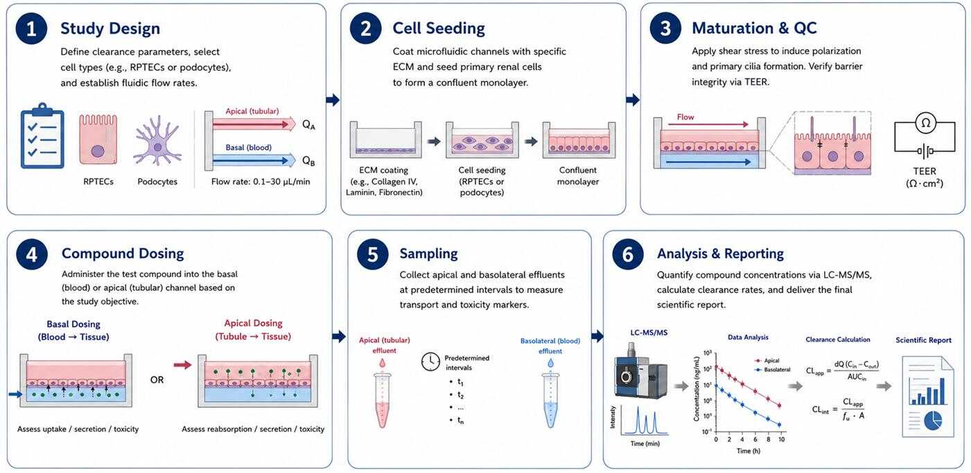
Service Workflow
Scientific Advantages of the Platform
- Hemodynamic shear stress restores in vivo-like expression of OAT and OCT transporters.
- Compartmentalization enables separate sampling from the basal (blood) and apical (urine) channels.
- Provides higher sensitivity for detecting early drug-induced kidney injury compared to 2D cultures.
FAQs
Why is fluidic shear stress necessary for renal models?
Shear stress is a critical biomechanical cue. It drives RPTECs to form primary cilia and microvilli, and ensures the physiological expression of influx and efflux renal transporters, which are typically lost in static cultures.
Which renal transporters are validated in this model?
Functional activity is validated for major basolateral uptake transporters (OAT1, OAT3, OCT2) and apical efflux transporters (MATE1, MATE2-K, P-gp, MRP2/4) utilizing specific fluorescent substrates and reference inhibitors.
Can the platform differentiate between apical and basolateral toxicity?
Yes, the dual-channel microfluidic design allows for independent administration and fluid sampling. We can determine if toxicity is driven by systemic exposure (basal channel) or tubular reabsorption (apical channel).
Is it possible to measure intracellular drug accumulation?
Yes. Cells can be lysed post-experiment to measure intracellular drug concentrations using LC-MS/MS. This is critical for evaluating accumulation-driven nephrotoxicities, such as those caused by aminoglycosides or platin-based compounds.
Initiate Your Renal Clearance Study
Consult with our scientific team to define flow rates, cell sources, and endpoints for your specific renal assays. We provide structured protocols tailored to your study design.
Online Inquiry