
Ready-to-use 3D human retinal organoids that recapitulate key retinal cell types and layered tissue organization for ophthalmology research, retinal disease modeling, and ocular drug discovery.
- Overview
- Details
- Advantages
- FAQs
Overview
The human retina is a highly specialized neural tissue responsible for converting light signals into visual information. It contains multiple interconnected cell populations organized into layered structures that are essential for visual processing and retinal homeostasis. Traditional 2D retinal cell cultures and animal models are often unable to fully reproduce the structural organization, cellular diversity, and developmental complexity of human retinal tissue.
Retinal organoids provide an advanced in vitro platform for studying retinal development, disease mechanisms, and therapeutic responses in a physiologically relevant human system. Derived from human induced pluripotent stem cells (hiPSCs), retinal organoids self-organize into three-dimensional retinal-like tissues containing multiple retinal cell types and layer-like organization.
What Are Retinal Organoids?
Retinal organoids are self-organizing 3D retinal tissue models generated from pluripotent stem cells under defined differentiation conditions. These organoids mimic key structural and functional features of the developing human retina and provide improved biological relevance compared with conventional monolayer cultures.
- Retina-like layered tissue organization
- Photoreceptor differentiation and maturation
- Multiple retinal neuronal cell populations
- Cell-cell and extracellular matrix interactions
- Long-term culture capability for retinal maturation studies
- Human-relevant ocular microenvironment
Depending on culture conditions and maturation stages, retinal organoids may contain photoreceptors, retinal ganglion cells, bipolar cells, Müller glia, and retinal pigment epithelium-associated cell populations relevant to retinal biology and disease research.
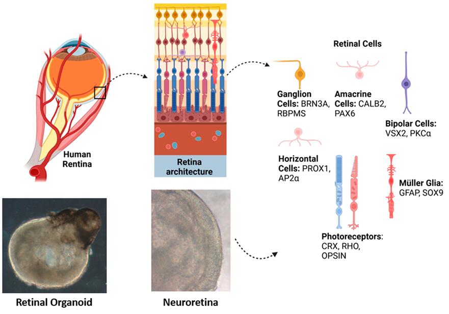 Fig. 1. Schematic overview of retinal organoid generation and ophthalmology research applications.
Our Ready-to-Use Retinal Organoids
Our retinal organoids are generated using optimized hiPSC differentiation and 3D culture technologies to produce reproducible retinal tissue models suitable for ophthalmology research, retinal disease modeling, and translational therapeutic development.
Key Features
- Human-relevant retinal tissue model. Recapitulates important structural and developmental features of human retinal tissue.
- Retina-like organization. Forms layer-like retinal architecture with multiple retinal-associated cell populations.
- Photoreceptor differentiation. Supports the development of rod- and cone-associated photoreceptor cell populations.
- Multicellular complexity. Contains retinal neuronal and supporting cell types within a physiologically relevant 3D microenvironment.
- Ready-to-use format. Cryopreserved organoids with optimized recovery protocols reduce preparation time and improve workflow efficiency.
- Batch consistency. Manufactured using standardized production and quality control procedures to ensure reproducibility.
Characterization & Validation
Our retinal organoids undergo extensive molecular, structural, and functional characterization to ensure biological relevance and experimental consistency.
- Retinal marker expression: Expression of CRX, Recoverin, Rhodopsin, Opsin, CHX10, and RPE-associated markers confirmed by immunostaining and qPCR analysis.
- 3D tissue morphology: Retina-like layered organization with neural retinal structures and photoreceptor-associated regions.
- Functional assessment: Evaluation of calcium signaling, neural activity, and retinal maturation-related functionality.
- Quality control: High post-thaw viability, sterility testing, and batch reproducibility validation.
Applications
Our ready-to-use retinal organoids support a broad range of ophthalmology and translational research applications.
- Retinal Disease Modeling: Study inherited retinal disorders, retinitis pigmentosa, age-related macular degeneration, glaucoma, and retinal degeneration using human-relevant retinal tissue systems.
- Ocular Drug Discovery: Evaluate drug efficacy, retinal toxicity, and therapeutic responses in physiologically relevant retinal tissue models.
- Retinal Development Research: Investigate retinal differentiation, photoreceptor maturation, retinal layering, and neurodevelopmental mechanisms.
- Personalized Medicine: Combine with patient-derived iPSC technologies for individualized disease modeling and therapeutic screening.
- Regenerative Medicine Research: Support studies related to retinal regeneration, cell replacement therapy, and tissue engineering.
Why Choose Our Retinal Organoids
- Physiologically relevant 3D retinal tissue model with retina-like organization
- Human-derived retinal system that improves translational relevance compared with animal models
- Supports photoreceptor development and retinal maturation studies
- Ready-to-use cryopreserved format saves weeks of differentiation and optimization time
- Reproducible production with standardized quality control procedures
- Compatible with multiple downstream assays including imaging, molecular analysis, and functional studies
- Versatile platform for retinal disease modeling, ocular drug screening, and gene therapy research
FAQs
Q: How are retinal organoids shipped?
Retinal organoids are supplied as cryopreserved samples and shipped on dry ice or in liquid nitrogen vapor shippers to maintain optimal viability and structural integrity during transportation.
Q: What retinal cell types are present in the organoids?
Depending on the differentiation stage and culture conditions, retinal organoids may contain photoreceptors, retinal ganglion cells, bipolar cells, Müller glia, and other retinal-associated cell populations.
Q: Are retinal organoids suitable for long-term culture?
Yes. Under optimized culture conditions, retinal organoids can be maintained long-term to support retinal maturation, disease modeling, and chronic treatment studies.
Q: Can retinal organoids be used for gene therapy studies?
Yes. Retinal organoids are compatible with gene delivery and gene editing workflows and are widely used in preclinical gene therapy and translational ophthalmology research.
Advance your ophthalmology research with physiologically relevant human retinal organoids.
Contact us today to request detailed product information, technical support, or customized retinal organoid solutions tailored to your research needs.
Online Inquiry