
Patient-derived lung cancer organoids that retain histological structure and key molecular features of primary lung tumors, supporting studies of tumor biology, drug response, and resistance mechanisms.
- Overview
- Details
- Advantages
- FAQs
Overview
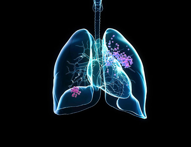
Lung cancer is one of the leading causes of cancer-related mortality worldwide. It is broadly classified into non-small cell lung cancer (NSCLC) and small cell lung cancer (SCLC), with NSCLC accounting for the majority of cases. Major NSCLC subtypes include lung adenocarcinoma and squamous cell carcinoma.
Lung tumors are highly heterogeneous at the molecular level, with frequent alterations in EGFR, KRAS, ALK, ROS1, BRAF, MET, and TP53. These genetic differences are closely associated with variability in disease progression and treatment response, particularly to targeted therapies and immune checkpoint inhibitors.
Conventional 2D cell lines often fail to reflect the complexity of primary tumors, especially in terms of architecture and intratumoral heterogeneity. Patient-derived lung cancer organoids provide a more representative model that preserves key disease features and supports translational research applications.
What Pathological Features Do Lung Cancer Organoids Recapitulate?
- Histological characteristics of NSCLC subtypes, including adenocarcinoma and squamous cell carcinoma
- Driver mutations in EGFR, KRAS, ALK, ROS1, BRAF, MET, and TP53
- Molecular subtype features associated with targeted therapy response
- Expression of lung cancer markers such as TTF-1, Napsin A, CK5/6, and EPCAM
- Patient-specific sensitivity to EGFR-TKIs, ALK inhibitors, and chemotherapy
- Tumor cell heterogeneity and clonal diversity
- Proliferation and invasion-associated phenotypes
- Immune-related signaling features relevant to immunotherapy research
These characteristics make lung cancer organoids suitable for studying tumor biology and evaluating therapeutic strategies in a patient-relevant system.
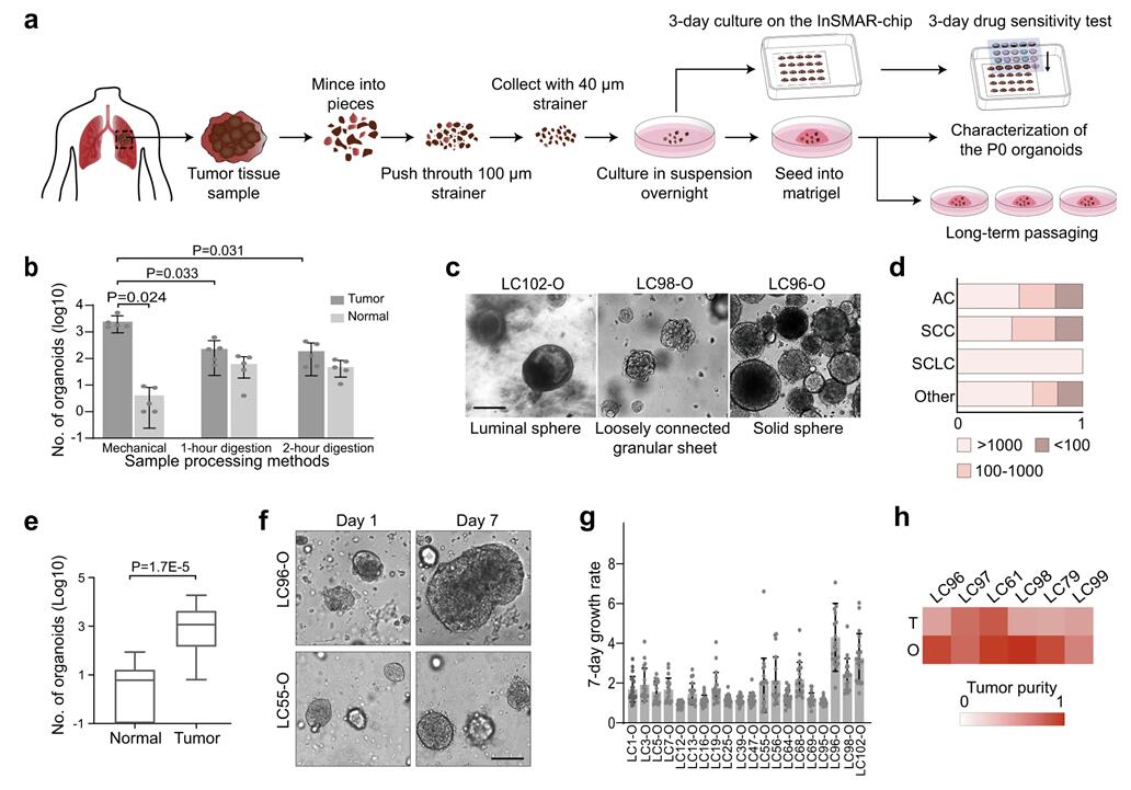 Fig. 1. Generation of lung cancer organoids (LCOs) from lung tumor tissues (Hu Y, Shui X, et al.,
2021).
Fig. 1. Generation of lung cancer organoids (LCOs) from lung tumor tissues (Hu Y, Shui X, et al.,
2021).
Our Lung Cancer Organoids
Our lung cancer organoids are derived from primary patient tumor tissues and maintained under optimized conditions to preserve clinically relevant molecular and histological characteristics.
Disease-Relevant Features
- Patient-derived models representing NSCLC and major histological subtypes
- Molecular heterogeneity reflecting key oncogenic driver alterations
- Preservation of tumor architecture consistent with primary lung tumors
- Clinically relevant biomarker expression for translational studies
- Cryopreserved, ready-to-use format for experimental workflows
Characterization & Validation
Each lung cancer organoid line is systematically characterized to ensure identity and disease relevance.
- Genetic profiling: Detection of driver mutations in EGFR, KRAS, ALK, ROS1, BRAF, MET, and TP53
- Biomarker validation: Assessment of TTF-1, Napsin A, CK5/6, EPCAM, and other markers
- 3D morphology assessment: Evaluation of organoid structure and growth patterns
- Disease phenotype analysis: Drug response and pathway activity profiling
- Quality control: Post-thaw viability ≥85%, identity confirmation, low batch variability, mycoplasma-free status
Applications
Lung cancer organoids provide a patient-relevant system to study tumor behavior under defined experimental conditions, especially in the context of driver mutations and therapy response variability.
- Oncogenic driver analysis: Explore functional consequences of EGFR, KRAS, ALK and other alterations in a 3D tumor context.
- Therapy response profiling: Evaluate sensitivity to targeted agents such as EGFR-TKIs, ALK inhibitors, and emerging combination regimens.
- Tumor evolution modeling: Study clonal dynamics and adaptation under long-term drug exposure.
- Immuno-oncology research: Support co-culture or conditioned systems for investigating tumor–immune signaling and immunotherapy-related mechanisms.
- Translational biomarker work: Link molecular features with phenotypic drug response to support biomarker identification and stratification strategies.
Why Choose Our Lung Cancer Organoids
- Patient-derived models that reflect clinical tumor heterogeneity
- Driver mutation–relevant systems for targeted therapy studies
- Ready-to-use format for rapid experimental setup
- Consistent quality supported by standardized validation
- Compatible with screening platforms for drug evaluation and translational research
FAQs
Q: Which lung cancer subtypes are included?
Models may include lung adenocarcinoma and squamous cell carcinoma, depending on the donor source.
Q: Are driver mutations characterized?
Yes. Key alterations such as EGFR, KRAS, ALK, ROS1, BRAF, MET, and TP53 can be profiled depending on the model.
Q: What assays are compatible?
The organoids support drug screening, imaging, immunostaining, gene expression profiling, and genomic analysis.
Q: Can customized lung cancer organoids be developed?
Yes. Custom models can be generated based on specific genetic backgrounds or research requirements.
Accelerate lung cancer research with physiologically relevant, patient-derived organoid models.
Contact us today to request detailed characterization data, pricing information, or customized lung cancer organoid solutions tailored to your research needs.
Online Inquiry