
Ready-to-use 3D human lung organoids that model airway and alveolar biology for respiratory disease research, drug screening, and infection studies
- Overview
- Details
- Advantages
- FAQs
Overview
The human lung is a complex organ responsible for gas exchange, host defense, and environmental sensing. Conventional in vitro models, such as 2D airway epithelial cells, and animal models often fail to fully capture the structural complexity and human-specific responses of lung tissue. This creates a need for more physiologically relevant in vitro models in respiratory research.
Lung organoids are advanced 3D human lung models derived from stem cells that self-organize into airway- or alveolar-like structures. These human lung organoids recapitulate key aspects of lung development, epithelial differentiation, and functional responses, providing a valuable platform for studying respiratory diseases and evaluating therapeutic candidates.
What Are Lung Organoids?
Lung organoids are self-organizing, multicellular 3D structures derived from human induced pluripotent stem cells (hiPSCs) or adult stem cells. They mimic essential features of lung tissue, including:
- 3D architecture resembling airway or alveolar compartments
- Presence of relevant epithelial cell types (e.g., ciliated cells, club cells, basal cells, type I and type II pneumocytes)
- Expression of lung-specific markers and functional proteins
- Capability for differentiation and maintenance under defined culture conditions
Depending on differentiation protocols, lung organoids can represent airway organoids or alveolar organoids, supporting diverse experimental applications in pulmonary biology.
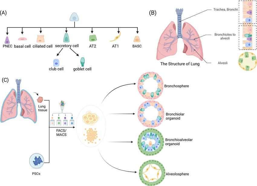 Fig. 1. The structure of lung and lung organoid (Huo Y, He S, et al., 2025).
Our Ready-to-Use Lung Organoids
Our human lung organoids are developed under controlled 3D culture conditions to support reproducible and biologically relevant outcomes. These ready-to-use lung organoids are suitable for respiratory disease modeling, drug screening, and infection studies.
Key Features
- Physiologically relevant models expressing lung markers such as NKX2.1, EPCAM, SPC (SFTPC), and MUC1
- Airway and alveolar differentiation enabling modeling of different lung regions
- Functional epithelial properties including mucus production and surfactant-related activity (depending on subtype)
- Cryopreserved and ready-to-use to streamline experimental workflows
Characterization & Validation
Each batch of lung organoids is characterized using molecular and functional assays to ensure consistency and relevance.
- Marker expression: NKX2.1, EPCAM, SFTPC, FOXJ1 validated by immunostaining and qPCR
- 3D morphology: Spherical or budding structures with organized epithelial layers
- Functional readouts: Cilia formation (airway models), surfactant-related protein expression (alveolar models)
- Quality control: Post-thaw viability ≥85%, low batch variability, mycoplasma-free
Applications
Our lung organoids support a range of applications in respiratory research and drug development:
- Disease Modeling: Study respiratory diseases such as asthma, COPD, pulmonary fibrosis, and lung cancer
- Drug Screening: Evaluate drug efficacy and pulmonary toxicity in human-relevant systems
- Infectious Disease Research: Investigate host–pathogen interactions, including viral infections (e.g., influenza, coronaviruses)
- Precision Medicine: Support patient-derived organoid research for personalized therapeutic evaluation
Why Choose Our Lung Organoids
- Human-relevant lung models reflecting airway and alveolar biology
- Ready-to-use format reduces time for model establishment
- Reproducible performance with standardized production and QC
- Flexible applications across respiratory disease models and drug screening
- Scalable for medium- to high-throughput experimental workflows
FAQs
Q: How are lung organoids delivered?
Lung organoids are shipped cryopreserved on dry ice or in liquid nitrogen. Standard thawing and recovery protocols allow rapid experimental setup.
Q: What types of lung organoids are available?
Our platform supports both airway organoids and alveolar-like organoids, depending on differentiation conditions and application needs.
Q: Can lung organoids be used for infection studies?
Yes. Lung organoids are suitable for studying respiratory pathogens and host responses in a controlled human-relevant system.
Q: How long can lung organoids be cultured after thawing?
They are typically used within 3–7 days for most assays but can be maintained longer under optimized culture conditions.
Enhance your respiratory research with reliable, ready-to-use human lung organoids.
Contact us for technical datasheets, pricing, or customized lung organoid solutions tailored to your project.
Online Inquiry