
Patient-derived melanoma organoids that faithfully recapitulate the cellular heterogeneity, mutational landscape, and drug response profiles of human cutaneous and metastatic melanoma.
- Overview
- Details
- Advantages
- FAQs
Overview
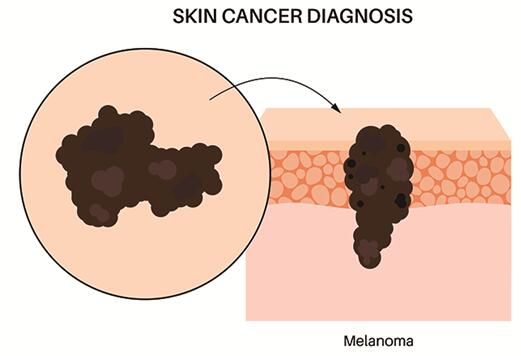
Melanoma is one of the most aggressive skin malignancies, characterized by high metastatic potential, rapid progression, and marked therapeutic resistance. Although targeted therapies and immunotherapies have improved outcomes in selected patients, intrinsic and acquired resistance remains a major clinical challenge.
Genomically, melanoma is defined by a high mutational burden and frequent alterations in the MAPK and PI3K signaling pathways, most notably BRAF, NRAS, and NF1 mutations. Additional changes in cell cycle regulators, immune checkpoint molecules, and epigenetic modifiers further contribute to tumor evolution and treatment failure.
Conventional 2D melanoma cell cultures often lose key phenotypic traits during long-term passage. In contrast, patient-derived melanoma organoids retain the architectural complexity, pigmentation patterns, and molecular diversity of primary and metastatic lesions, providing a robust platform for translational research.
Key Pathological Features Recapitulated by Melanoma Organoids
- Histological resemblance to primary and metastatic melanoma
- Frequent driver mutations in BRAF, NRAS, and NF1
- Melanocytic differentiation markers including SOX10, MITF, and S100
- Variable pigmentation and morphological heterogeneity
- Elevated tumor mutational burden and neoantigen diversity
- Resistance profiles to BRAF/MEK inhibitors and immune checkpoint blockade
- Invasive and metastatic phenotypes in 3D culture systems
- Preservation of patient-specific immune microenvironment features when co-cultured
These characteristics make melanoma organoids an ideal model for studying tumor evolution, drug response, and resistance mechanisms in a clinically relevant context.
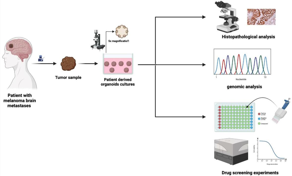 Fig. 1. Melanoma Brain Metastases Patient-Derived Organoids (Abedellatif S-E, Hosni R, et al., 2024).
Our Melanoma Organoids
Our melanoma organoids are derived from patient tumor tissues and propagated under optimized, serum-free conditions to maintain phenotypic stability, genetic integrity, and functional relevance.
Disease-Relevant Features
- Patient-derived models representing cutaneous, acral, mucosal, and metastatic melanoma
- Preserved melanocytic lineage identity with physiological pigmentation patterns
- Mutational diversity across MAPK and PI3K signaling pathways
- Tumor heterogeneity reflecting inter- and intra-patient variation
Characterization & Quality Assurance
Each melanoma organoid line is comprehensively validated to ensure biological fidelity and research-grade quality.
- Genetic profiling: Mutation analysis of BRAF, NRAS, NF1, and other melanoma-associated genes
- Immunophenotyping: Expression of SOX10, MITF, S100, and melanoma differentiation antigens
- Morphological assessment: 3D structure, pigmentation, and growth kinetics
- Functional validation: Drug sensitivity profiling against targeted and immunotherapy agents
- Quality control: Post-thaw viability ≥85%, identity confirmation, mycoplasma-free certification
Applications
Melanoma organoids enable a wide range of preclinical and translational investigations:
- Targeted Therapy Research: Evaluation of BRAF, MEK, and combinatorial inhibition strategies.
- Immuno-Oncology: Modeling tumor–immune interactions and checkpoint blockade responses.
- Resistance Mechanisms: Dissection of adaptive and acquired resistance pathways.
- Precision Medicine: Patient-specific drug response prediction.
- Metastasis Studies: Analysis of invasive behavior and organotropic colonization.
Why Choose Our Melanoma Organoids
- Clinically Relevant Models: Derived from real patient tumors across melanoma subtypes
- Genomic & Phenotypic Fidelity: Preserve key mutations and lineage-specific traits
- Immune-Compatible: Amenable to co-culture with immune cells and fibroblasts
- Translational Utility: Ideal for drug discovery, biomarker validation, and precision oncology
FAQs
Q: How long does it take to establish a new melanoma organoid line from patient tissue?
Typical establishment timelines range from 4 to 8 weeks, depending on tumor cellularity, sample quality, and the selected culture conditions. Expedited protocols are available for time-sensitive projects.
Q: Are melanoma organoids suitable for high-throughput drug screening?
Yes. Our cryopreserved melanoma organoids are optimized for 96- and 384-well plate formats, supporting large-scale compound screening, combination studies, and dose–response profiling.
Q: Can I use these organoids for multi-omics studies?
Absolutely. Melanoma organoids are compatible with genomics, transcriptomics, proteomics, and single-cell sequencing workflows, enabling deep molecular characterization alongside phenotypic assays.
Q: Do you provide matched normal and tumor organoid pairs?
In select cases, we can generate matched normal melanocyte and tumor organoid pairs from the same donor, facilitating comparative studies on oncogenic transformation and drug selectivity.
Q: How stable are melanoma organoids during long-term culture?
With our optimized, serum-free culture system, melanoma organoids maintain stable morphology, mutational profiles, and drug response characteristics over extended passaging, ensuring consistency across experiments.
Q: What melanoma subtypes are represented in your organoid collection?
Our library includes cutaneous, acral, mucosal, and metastatic melanoma organoids, covering the major clinical subtypes encountered in translational research.
Empower melanoma research with patient-derived organoid models that bridge molecular discovery and clinical translation.
Contact us to request detailed characterization data, pricing, or custom melanoma organoid solutions tailored to your research objectives.
Online Inquiry