
Ready-to-use human esophageal organoids that recapitulate stratified squamous epithelium for disease modeling, drug screening, and epithelial biology research
- Overview
- Details
- Advantages
- FAQs
Overview
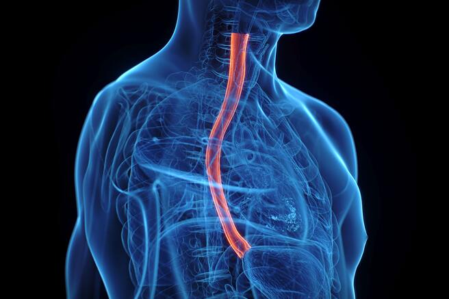
The human esophagus is lined by a stratified squamous epithelium that serves as a protective barrier against mechanical stress, chemical exposure, and pathogens. Traditional 2D epithelial cultures and animal models are limited in their ability to reproduce the multilayered structure, differentiation gradients, and barrier function of human esophageal tissue. This creates a need for advanced in vitro systems that more accurately reflect human physiology.
Esophageal organoids are 3D multicellular models derived from human stem cells that self-organize into structures resembling native esophageal epithelium. These organoids recapitulate key features such as basal progenitor layers and differentiated suprabasal cells, providing a biologically relevant platform for studying epithelial homeostasis and disease.
What Are Esophageal Organoids?
Organoids are self-organizing 3D structures that mimic essential aspects of organ architecture and function. Esophageal organoids exhibit:
- Stratified squamous epithelial organization
- Presence of basal stem/progenitor cells and differentiated epithelial layers
- Barrier-associated functions and response to environmental stimuli
- Capacity for expansion and controlled differentiation in culture
These features make them highly suitable for investigating esophageal biology and disease mechanisms in a human-relevant context.
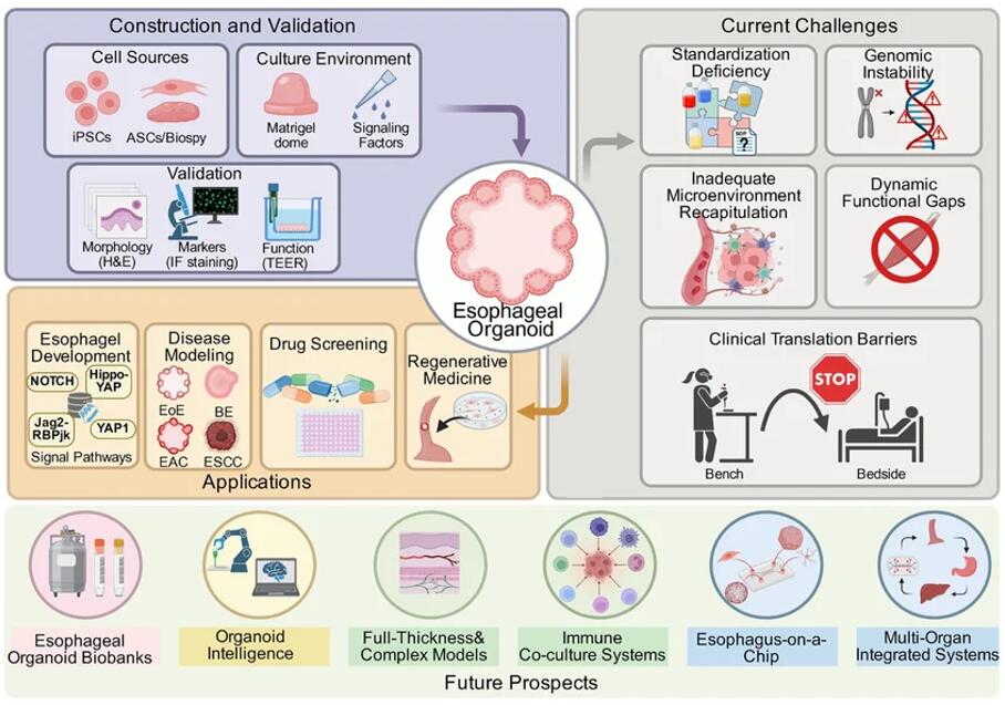 Fig. 1.
Overview of esophageal organoid construction, applications, current challenges, and future prospects (An J, en
W, et al., 2026).
Fig. 1.
Overview of esophageal organoid construction, applications, current challenges, and future prospects (An J, en
W, et al., 2026).
Our Ready-to-Use Esophageal Organoids
Our esophageal organoids are generated from human induced pluripotent stem cells (hiPSCs) using optimized protocols that promote stratified epithelial differentiation and structural organization. Delivered in a cryopreserved, ready-to-use format, these organoids enable rapid experimental setup with high reproducibility.
Key Features
- Physiological relevance: Mimic stratified squamous epithelium of the human esophagus
- Cellular diversity: Include basal progenitor and differentiated epithelial cell populations
- High consistency: Controlled production ensures reproducible morphology and function
- Ready-to-use: Cryopreserved for convenient recovery and immediate use
Characterization & Validation
Our esophageal organoids undergo rigorous validation to ensure biological fidelity and experimental reliability.
- Marker expression: p63, KRT5 (basal cells), KRT13 and involucrin (differentiated layers)
- 3D morphology: Stratified epithelial architecture with clear layer organization
- Functional properties: Barrier-related responses and epithelial differentiation capacity
- Quality control: ≥85% post-thaw viability, mycoplasma-free, low batch variability
Applications
Esophageal organoids serve as a versatile platform for multiple research areas:
- Disease Modeling: Study esophageal diseases such as Barrett's esophagus, esophagitis, and esophageal cancer
- Drug Screening: Evaluate therapeutic efficacy and epithelial toxicity
- Barrier Function Studies: Investigate epithelial integrity and response to chemical or mechanical stress
- Regenerative Research: Explore epithelial repair and stem cell-driven regeneration
Why Choose Our Esophageal Organoids
- Human-relevant epithelial model with stratified structure
- Ready-to-use format reduces experimental preparation time
- Reproducible and consistent across batches
- Scalable for high-throughput screening applications
- Broad applicability in disease, toxicity, and barrier studies
FAQs
Q: How are esophageal organoids shipped?
They are shipped in cryopreserved vials on dry ice or in liquid nitrogen to ensure optimal viability upon delivery.
Q: How long can they be cultured after thawing?
They are typically used within 3–7 days for most assays, with longer culture possible under optimized conditions.
Q: Are these organoids suitable for barrier function studies?
Yes, their stratified epithelial structure makes them ideal for studying epithelial integrity and barrier responses.
Q: Do you offer disease-specific esophageal organoids?
Currently, we offer normal esophageal organoids. Disease-specific and genetically engineered models are under development and available upon request.
Advance esophageal research with physiologically relevant, ready-to-use human esophageal organoids.
Contact us to request datasheets, pricing, or customized solutions tailored to your research needs.
Online Inquiry