
Patient-derived bladder cancer organoids that retain key molecular and histological features of human urothelial tumors, supporting studies of disease biology, drug response, and biomarker development.
- Overview
- Details
- Advantages
- FAQs
Overview
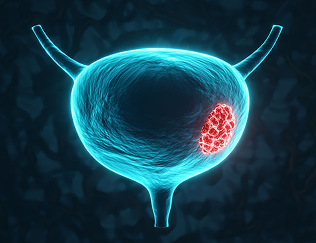
Bladder cancer is one of the most common malignancies of the urinary system. The majority of cases are urothelial carcinomas, which include non-muscle-invasive and muscle-invasive disease. Despite available surgical and systemic treatments, recurrence and progression remain frequent clinical challenges.
Bladder tumors are molecularly heterogeneous, with frequent alterations in FGFR3, TP53, RB1, PIK3CA, ERBB2, and TERT promoter regions. These alterations contribute to differences in tumor behavior and treatment response across patients. Conventional 2D models often fail to reproduce this diversity.
Our bladder cancer organoids are derived from patient tumor tissues and maintain key histological and molecular features of the original tumors. These models are suitable for studying tumor biology, drug response, resistance mechanisms, and biomarker discovery.
What Pathological Features Do Bladder Cancer Organoids Recapitulate?
- Histological characteristics of urothelial carcinoma
- Common mutations including FGFR3, TP53, RB1, PIK3CA, and ERBB2
- Luminal and basal subtype-associated features
- Expression of markers such as GATA3, CK5, CK20, UPK, and EPCAM
- Patient-specific drug response profiles
- Tumor heterogeneity observed in primary bladder cancer
- Proliferation and invasion-related phenotypes
- Clinically relevant response patterns to therapies
These characteristics make bladder cancer organoids suitable for translational research and preclinical evaluation.
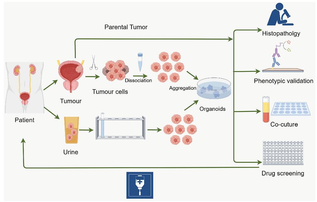 Fig. 1. Establishment of BCOs and the process for precision medicine applications (Zheng Q, Liao E,
et al., 2026).
Fig. 1. Establishment of BCOs and the process for precision medicine applications (Zheng Q, Liao E,
et al., 2026).
Our Bladder Cancer Organoids
Our bladder cancer organoids are generated from patient-derived tumor tissues and maintained under defined culture conditions to preserve disease-relevant characteristics.
Disease-Relevant Features
- Patient-derived models reflecting primary bladder tumor characteristics
- Molecular subtype representation including luminal and basal features
- Genetically relevant profiles with common bladder cancer-associated alterations
- Preserved tumor heterogeneity across patient samples
- Cryopreserved format suitable for immediate experimental use
Characterization & Validation
Each organoid batch is characterized to confirm identity, quality, and disease relevance.
- Genetic profiling: Detection of key bladder cancer-associated mutations
- Biomarker validation: Analysis of GATA3, CK5, CK20, UPK, and EPCAM expression
- 3D morphology assessment: Evaluation of organoid structure and growth behavior
- Disease phenotype evaluation: Drug response and proliferation profiling
- Quality control: Post-thaw viability ≥85%, identity verification, low batch variation, mycoplasma-free status
Applications
These organoids are suitable for multiple areas of bladder cancer research:
- Disease Biology: Study tumor development and molecular subtype differences.
- Drug Screening: Evaluate chemotherapy, targeted agents, and combination therapies.
- Precision Medicine: Assess patient-specific treatment responses.
- Resistance Studies: Investigate mechanisms of therapy resistance.
- Biomarker Research: Identify and validate predictive biomarkers.
Why Choose Our Bladder Cancer Organoids
- Patient-derived models reflecting clinical tumor features
- Subtype-relevant representation of bladder cancer heterogeneity
- Ready-to-use format for streamlined workflows
- Consistent quality supported by standardized validation
- Screening-compatible for drug evaluation studies
FAQs
Q: What types of bladder cancer are represented?
Models may represent non-muscle-invasive and muscle-invasive urothelial carcinoma depending on the donor tissue.
Q: Which biomarkers are available for validation?
Common bladder cancer markers such as GATA3, CK5, CK20, UPK, and EPCAM can be assessed.
Q: What assays are supported?
Applications include viability assays, imaging, immunostaining, gene expression analysis, and drug screening.
Q: Can custom models be developed?
Yes. Custom bladder cancer organoid models can be generated based on specific research requirements.
Accelerate bladder cancer research with physiologically relevant, ready-to-use human tumor organoids.
Contact us today to request detailed characterization data, pricing information, or customized bladder cancer organoid solutions tailored to your research needs.
Online Inquiry