Unlock breakthroughs in neurodegenerative disease research with human-relevant brain organoids and blood-brain barrier-on-a-chip platforms for modeling disease pathology, drug screening, and personalized therapeutic development.
- Overview
- Platform
- Service
- Application
- Workflow
- FAQs
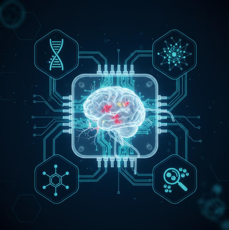
Neurodegenerative disorders, including Alzheimer's disease, Parkinson's disease, Amyotrophic Lateral Sclerosis (ALS), and Huntington's disease, are characterized by the progressive loss of structure and function of neurons, leading to cognitive decline, motor dysfunction, and ultimately, a profound impact on quality of life. The complexity of the human brain, the involvement of multiple cell types (neurons, astrocytes, microglia, oligodendrocytes), and the selective vulnerability of specific neuronal populations have made these diseases notoriously difficult to model accurately. Traditional animal models and 2D neuronal cultures fail to fully recapitulate the human brain's 3D architecture, cellular diversity, and the complex, non-cell autonomous interactions that drive disease progression.
Brain organoids and neural organ-on-a-chip systems represent a paradigm shift. Cerebral organoids derived from patient-specific iPSCs can self-organize into 3D structures containing region-specific neuronal subtypes, functional neural networks, and supporting glial cells, modeling key pathological hallmarks like amyloid-beta plaques, tau tangles, and alpha-synuclein aggregates. Complementing this, blood-brain barrier (BBB)-on-a-chip models incorporate endothelial cells, pericytes, and astrocytes under physiological flow to accurately predict drug penetration into the brain, a major hurdle in CNS drug development. Together, these platforms offer unprecedented opportunities to study disease mechanisms, screen for neuroprotective compounds, and develop personalized treatment strategies.
Comparison of Models for Neurodegenerative Disease Research
| Model Type | Recapitulation of Brain Complexity | Human Genetic & Cellular Relevance | Suitability for High-Content Screening | Ability to Model Cell-Cell Interactions (e.g., Neuroinflammation) |
| 2D Neuronal Culture | Low; lacks 3D cytoarchitecture and network complexity. | Moderate (if human iPSC-derived) | High | Limited; difficult to co-culture multiple CNS cell types effectively. |
| Animal Models (Transgenic Rodents) | Moderate; captures some systemic and behavioral aspects. | Low; significant species differences in brain physiology and disease mechanisms. | Very Low | High, but murine immune system differs from human. |
| Post-Mortem Human Brain Tissue | High for end-stage pathology. | Highest (direct human tissue) | None | Static snapshot; cannot model disease progression or intervention. |
| Brain Region-Specific Organoids | High; 3D structure, multiple neuronal layers, and some glial cells. | Highest (patient iPSC-derived) | Medium to High | Good; can incorporate microglia and astrocytes to model neuroinflammation. |
| Assembloids (Fused Organoids) | Very High; models circuit formation between brain regions (e.g., cortex-striatum). | Highest | Medium | Excellent; ideal for studying connectivity and circuit-based pathologies. |
| BBB-on-a-Chip & Multi-organ CNS Chips | Specialized; models the neurovascular unit and systemic interactions. | High (human cell-based) | Medium | Superior; explicitly models neuron-astrocyte-endothelial-immune crosstalk under flow. |
Our Neurodegenerative Disease Modeling Platforms: Brain Organoids & Neural MPS
We provide a cutting-edge suite of platforms featuring region-specific brain organoids, assembloids, and microphysiological systems (MPS) to model the complexity of the human central nervous system and its diseases.
Key Features:
- Comprehensive Brain Organoid Biobank: Access to a library of iPSC lines from patients with familial and sporadic forms of Alzheimer's, Parkinson's, ALS, Frontotemporal Dementia (FTD), and Huntington's disease, along with isogenic CRISPR-corrected controls.
- Region-Specific & Assembloid Models: Generate cerebral cortex, midbrain (dopaminergic), spinal cord, hippocampal, and striatal organoids. Create assembloids (e.g., cortical-striatal, cortical-thalamic) to model circuit dysfunction and axonal trafficking defects.
- Advanced BBB-on-a-Chip: A dynamic tri-culture model (brain microvascular endothelial cells, pericytes, astrocytes) under physiological shear stress for predictive assessment of CNS drug permeability and efflux transport.
- Functional & Molecular Readouts: Integrated multi-electrode arrays (MEAs) for network electrophysiology, calcium imaging for neuronal activity, high-content imaging for protein aggregation (e.g., phospho-tau, α-synuclein).
- Glial & Immune Integration: Seamlessly incorporate microglia (brain-resident immune cells) and peripheral immune cells to model neuroinflammation, a key driver of neurodegeneration.
Neurodegenerative Disease Research and Drug Discovery Services
Our end-to-end services leverage human-relevant models to de-risk and accelerate therapeutic development for neurodegenerative conditions.
- Disease Modeling & Phenotypic Characterization: Establish and deeply phenotype patient-derived or gene-edited brain organoid models. Quantify key hallmarks: protein aggregation (Aβ, p-tau, α-syn, TDP-43), neuronal loss, synaptic dysfunction, and neuroinflammatory responses.
- High-Content Neuroprotective Drug Screening: Screen small molecules, biologics, or ASOs in 96/384-well formatted organoid assays. Readouts include cell viability, reactive oxygen species, mitochondrial function, and clearance of pathological protein aggregates.
- Blood-Brain Barrier Penetration & PK/PD Studies: Utilize our BBB-on-a-chip to rank-order compound permeability (Papp), assess efflux transporter involvement (P-gp, BCRP), and predict brain exposure. Perform integrated BBB-organoid studies to measure functional efficacy post-penetration.
- Functional Genomics & Target Discovery: Conduct CRISPR knockout/aactivation screens in disease organoids to identify genetic modifiers and novel therapeutic targets. Validate targets via rescue experiments in isogenic backgrounds.
- Personalized Medicine & Clinical Trial Stratification: Generate a "patient-in-a-dish" model from an individual's iPSCs to test response to standard-of-care or novel therapies, informing treatment decisions and clinical trial enrollment.
Core Applications in Neurodegenerative Disease Research
Brain Organoids for Disease Mechanism & Therapy
- Alzheimer's Disease Modeling: Cerebral organoids that spontaneously develop extracellular amyloid-beta plaques and intracellular hyperphosphorylated tau tangles over extended culture, enabling the study of the amyloid-tau cascade and testing of anti-aggregation therapies.
- Parkinson's Disease & Lewy Body Pathology: Midbrain organoids containing dopaminergic neurons that develop α-synuclein aggregates (Lewy body-like inclusions), mitochondrial dysfunction, and selective neuronal vulnerability, ideal for testing neuroprotective agents.
- ALS/FTD & TDP-43 Proteinopathy: Cortical and spinal cord organoids that model TDP-43 mislocalization and aggregation, neuronal hyperexcitability, and glial toxicity, relevant for C9orf72 and other genetic forms.
Organ-on-a-Chip for Systemic & Translational Studies
- Blood-Brain Barrier (BBB) Disruption & Drug Delivery: Model the breakdown of the BBB in neuroinflammation or assess novel delivery strategies (nanoparticles, focused ultrasound) for enhanced CNS drug uptake. Measure trans-endothelial electrical resistance (TEER) and permeability in real-time.
- Gut-Brain Axis Modeling: A connected gut-chip and brain-organoid system to investigate the role of the microbiome, gut-derived metabolites, and peripheral inflammation in driving neuroinflammation and pathology in Parkinson's and Alzheimer's disease.
- Neurovascular Unit (NVU) on-a-Chip: Integrate neurons, astrocytes, and brain microvascular endothelial cells to study the critical role of vascular dysfunction and hypoperfusion in neurodegenerative processes, and test interventions that improve cerebral blood flow.
Workflow for Neurodegenerative Disease Modeling & Screening
iPSC Line Establishment & Characterization
Generate or source patient-specific iPSCs. Perform rigorous QC for pluripotency, karyotype, and identity. Create isogenic controls via gene editing if required.
Brain Organoid/
Assembloid Differentiation
Directed differentiation into region-specific organoids (cortical, midbrain, spinal cord) or assembloids using optimized, reproducible protocols over 8-12 weeks.
Phenotypic Validation & Disease Staging
Comprehensive characterization: Immunostaining for cell-type markers and pathological proteins, transcriptomics, electrophysiology (MEA), and functional assays to confirm disease phenotype emergence.
Assay Development & Compound Screening
Develop robust, miniaturized HTS/HCS assays (e.g., imaging-based aggregation, neuronal activity, cytotoxicity). Perform screening of compound libraries in 96/384-well format.
Integrated Analysis & Hit Validation
Multi-parametric data analysis to identify hits. Validate top candidates in secondary assays (e.g., BBB penetration, efficacy in isogenic controls, dose-response) and mechanistic studies.
FAQs
How long does it take to generate mature brain organoids that show disease pathology?
Timelines vary by disease and protocol. Cortical organoids can show early neuronal network activity by 8-10 weeks. For pathologies like amyloid-beta plaques or tau tangles in Alzheimer's models, extended maturation of 3-6 months or the use of pro-aging factors may be required to accelerate phenotype development. We offer both standard and accelerated protocols.
Can you model neuroinflammation using these platforms?
Yes. This is a key strength. We can:
- Innate Immunity: Integrate iPSC-derived microglia into brain organoids to study their role in phagocytosis, cytokine release, and synaptic pruning.
- Peripheral Immunity: Introduce peripheral immune cells (e.g., T cells, monocytes) into BBB-on-a-chip models to study their migration and contribution to neuroinflammation.
- Readouts include cytokine profiling, phagocytosis assays, and imaging of microglial morphology.
What is the advantage of using assembloids over single-region organoids?
Assembloids (fused organoids) model neural circuits and long-distance connectivity, which is crucial for diseases where pathology spreads (e.g., tau in Alzheimer's, α-synuclein in Parkinson's) or where specific circuit dysfunction is central (e.g., cortical-striatal in Huntington's). They allow the study of axonal transport deficits, synaptic connectivity between regions, and network-level dysfunction that cannot be captured in isolated region models.
Online Inquiry